What Is EMG? A Beginner’s Guide to the Signal Itself (Part 1: The Physiological Origin)
Electromyography (EMG) is the electrical signal your muscles produce when they work. Every move you make, from blinking to lifting a weight, starts as an electrical event deep inside your body. This article explains where that signal comes from, how it is generated by motor neurons and muscle fibers, what shapes it into the waveforms we record, and how it changes as you contract harder. If you want to understand the raw material of EMG before any processing or filtering, read on.
The Physiological Origin: Where the Signal Comes From
Your brain and muscles speak the same language. That language is electricity and chemicals.
When we classify muscles as voluntary and involuntary, skeletal muscles are the voluntary ones. These are the muscles that let you walk, write, or lift a cup.
Skeletal muscles attach to bones. They bring together muscle fibers, connective tissue, blood vessels, and nerve endings.
Muscle fibers are long, cylindrical cells. They have multiple nuclei. These fibers group into bundles called fascicles.
The sarcolemma wraps around each muscle fiber. It is a specialized membrane that can transmit electrical signals.
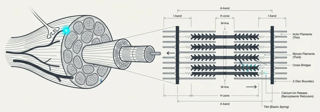
Inside the sarcolemma, you find myofibrils. Myofibrils contain thin actin and thick myosin filaments. These filaments slide over each other. ATP provides the energy for this movement. When they slide, the muscle shortens or lengthens. That is contraction and relaxation.
Like other excitable tissues that evolved in water, muscles contain ions that dissolve in water.
Between the sarcolemma and myofibrils sits the sarcoplasmic reticulum. It stores calcium ions and releases them when needed for contraction. When calcium comes out, a protein called troponin grabs it. That triggers contraction.
Sodium and potassium ions handle a different job. They create and transmit action potentials. Without them, contraction never starts.
The inside of a resting muscle fiber is more negative than the outside. This happens because of negatively charged molecules trapped inside the cell. Proteins and RNA both carry negative charges, and they cannot cross the membrane. At the same time, the membrane lets potassium ions pass through more easily than sodium ions. Potassium flows out, taking positive charge with it.
This creates a steady voltage difference of about -70 millivolts between the inside and outside. Physiologists call this the resting membrane potential.
Muscle fibers have two special abilities. They can respond to an electrical signal. And they can pass that signal along their entire length.
But where do these electrical signals come from?
The Motor Neuron: Where the Signal Starts
The electrical signals that excite muscle fibers do not begin in the muscle. They begin in the spinal cord.
Motor neurons reside in the ventral horn. Some neurons, like the cranial nerves that control facial muscles, are located in the brain stem.
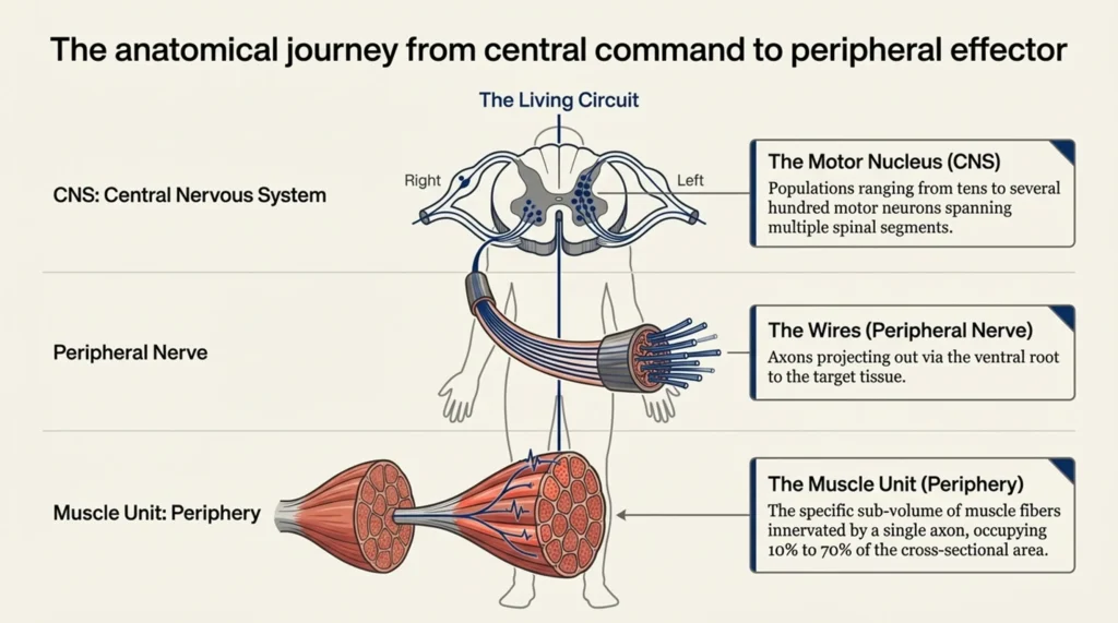
The anatomy of motor neurons consists of three parts. Dendrites are branched extensions that receive signals. The cell body integrates incoming signals. The axon is the part that carries the signal to the muscles.
Commands from the brain travel through multiple descending pathways including the corticospinal, rubrospinal, vestibulospinal, and reticulospinal tracts and reach motor neurons. Sensory feedback from sensory organs also converges on motor neurons. When the fluctuating extracellular voltage level around the neuron and the threshold that varies with the neuron’s own structure are exceeded, it generates an action potential. This signal is transmitted to the muscle fibers.
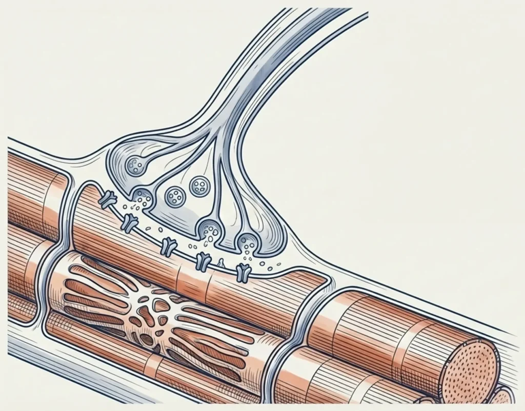
At the end of the axon, a small gap remains near the muscle fiber. This gap is called the synaptic cleft. When the action potential reaches the axon terminal, calcium channels open. Calcium enters and causes vesicles to release acetylcholine. Acetylcholine spreads across the synaptic cleft and binds to receptors on the muscle fiber membrane. This binding opens sodium channels. Sodium rushes in. The muscle fiber depolarizes. The chemical signal has turned back into an electrical signal.
The motor axon branches out as it enters the muscle. Each branch connects to a single muscle fiber. A single motor neuron can connect from a few fibers to thousands, depending on the muscle fiber type. One motor neuron and all the muscle fibers it connects to together are called a motor unit.
The Motor Unit: The Fundamental Functional Unit
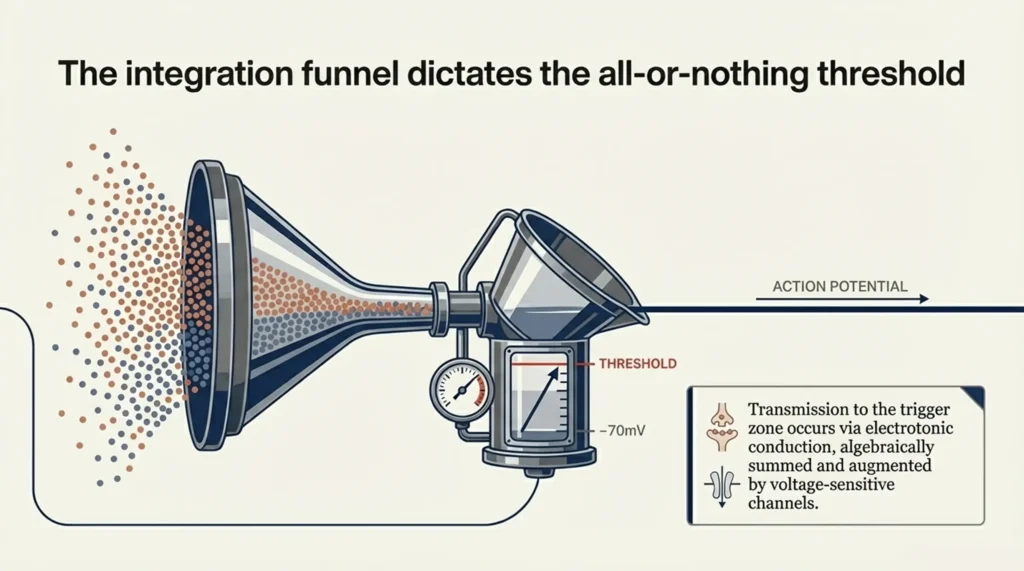
A motor unit works on an all or nothing principle.
When a motor neuron fires, all the muscle fibers it innervates contract together. There is no graded contraction. Either all of them fire or none of them fire.
Small motor units contain few muscle fibers. Examples include eye muscles and hand muscles. They coordinate fine movements.
Large motor units contain thousands of muscle fibers. Leg muscles are examples. Large motor units are used for powerful movements.
The muscle fibers of one motor unit are not grouped together in one place. They are intermingled with fibers from other motor units. This allows the muscle to contract smoothly and evenly.
During low force production, small motor units are recruited first. As force increases, larger motor units are also recruited. This is called Henneman’s size principle.
Why are small motor units recruited first? The answer lies in the motor neuron itself. Small motor neurons have higher input resistance, which means lower input conductance. They reach the firing threshold with less synaptic current. Large motor neurons have lower input resistance, which means higher input conductance. They need more synaptic current to fire, so they are recruited later.
The Action Potential: The Electrical Signal Itself
An action potential is a rapid reversal of the membrane voltage.
When a muscle fiber receives a strong enough signal at the neuromuscular junction, voltage-gated sodium channels open. Sodium ions rush into the cell. The inside of the fiber becomes positive, reaching about +30 millivolts. This phase is called depolarization.
Almost immediately, sodium channels close. Voltage-gated potassium channels open. Potassium ions flow out of the cell. The inside becomes negative again. This phase is called repolarization.
Sometimes the membrane overshoots slightly below the resting potential before stabilizing. This is called hyperpolarization.
The entire event lasts a few milliseconds. It follows the all or nothing principle. If the stimulus reaches threshold, the full action potential fires. If not, nothing happens.
Once generated, the action potential travels along the muscle fiber membrane. It propagates in both directions from the neuromuscular junction toward the ends of the fiber.
The speed of this propagation is called conduction velocity. It depends on fiber diameter. Larger fibers conduct faster.
The action potential is the trigger. It tells the sarcoplasmic reticulum to release calcium. Calcium starts the contraction. Without the action potential, the muscle fiber remains relaxed.
Every action potential that travels along a muscle fiber contributes to the electrical signal that electrodes can detect on the skin. This is where EMG begins.
The Motor Unit Action Potential (MUAP): The EMG Building Block
When a motor neuron fires, every muscle fiber in its motor unit generates its own action potential. These potentials do not all reach the electrode at the same time. They travel through the tissue, sum together, and form a waveform that is unique to that motor unit. This waveform is called the motor unit action potential, or MUAP.
With a needle electrode, you can separate individual MUAPs. But with surface electrodes, you measure the sum of many MUAPs, filtered by the electrode shape and the tissue.
Larger motor units produce larger MUAPs. The distance between each fiber and the electrode determines how much that fiber contributes to the MUAP. If a fiber is far from the needle electrode, its contribution is smaller and its potential looks smoother and wider.
How fibers are arranged inside the muscle, whether scattered or clustered, also shapes the MUAP.
A needle electrode sits inside the muscle. It records MUAPs that are sharp and clear. A surface electrode sits on the skin. The signal passes through tissue and gets filtered. That is why MUAPs in surface EMG look wider and smoother. Also, a surface electrode picks up MUAPs from many motor units at the same time.
At low force levels, MUAPs are sparse. As force increases, more MUAPs appear and the signal becomes dense. This density is not random noise. It creates what we call an interference pattern. Separating individual MUAPs from surface EMG becomes an engineering problem.
In the clinic, MUAPs help with diagnosis:
- In neuropathy, MUAPs are large and long.
- In myopathy, MUAPs are small, short, and often polyphasic.
- In ALS, you can see both types together.
From Rest to Contraction: How Electrical Activity Changes with Force
A resting muscle is electrically silent. But the moment you decide to move, everything changes.
At low force levels, only a few motor units are active. The first to appear are the small motor units. MUAPs are sparse.
As force increases, more motor units join in. First small, then medium, then large motor units are recruited. At the same time, the firing rate of already active motor units also increases. This is called rate coding.
When force gets very high, almost all motor units become active. At some point, there are no new motor units left to recruit. They are all already firing. To increase force further, the active motor units must fire faster. This is why rate coding becomes more dominant at maximal forces.
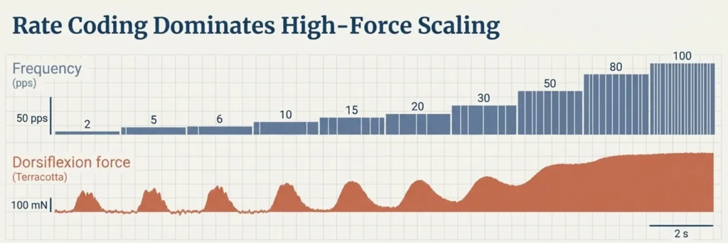
MUAPs overlap completely. The interference pattern becomes dense.
In general, as force increases, EMG amplitude also increases. But this relationship is not linear. It varies depending on the muscle type. Factors like fatigue, muscle length, and contraction speed all affect this relationship.
Recruitment dominates at low and moderate forces. Rate coding dominates at high forces. Together, they adjust the force produced by the muscle.
Further Reading
For a deeper understanding of the topics covered in this article, the following textbook is an excellent resource:
Merletti, R., & Farina, D. (Eds.). (2016). Surface Electromyography: Physiology, Engineering, and Applications. IEEE Press / John Wiley & Sons.
https://www.wiley.com/en-gb/-p-9781118987025
This is the first part of a series. In the next article, we will look at how we actually record this signal with electrodes and amplifiers.